RIMAL LAB
Research

Rimal Lab

Research

Research

Research programmes

Four concise programmes outline questions and methods to connect p53 transcriptional outputs (including DRAM1) to autophagy, organelle contacts, calcium signalling and cell-cycle outcomes.
01

DRAM1 and the p53–DRAM1 axis

How does p53-driven DRAM1 expression influence lysosomal function, organelle contacts and cell fate under genotoxic or mitochondrial stress?

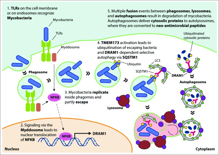
DRAM1 was identified as a p53-inducible lysosomal protein required for p53-mediated autophagy and apoptosis. Recent experimental work implicates DRAM1 in modulating organelle structure and inter-organelle contacts: excess DRAM1 can perturb ER morphology and trigger ER stress while promoting ER-phagy. Understanding these pleiotropic roles of DRAM1 is central to connecting p53 transcriptional programmes to cellular homeostasis.

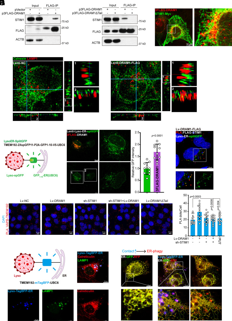
This programme explores DRAM1’s cellular localisation determinants, its interaction partners at lysosome–ER interfaces (for example STIM1), and the functional consequences for calcium homeostasis, ER stress signalling and cell survival. We will use genetic manipulation of DRAM1 expression combined with high-resolution imaging and functional assays of calcium and ER stress to map DRAM1 activities.
Key discovery linking p53-regulated DRAM1 to lysosome–ER contact and ER stress (figure from Wang et al., PNAS 2024).
02

Autophagy (canonical and alternative) in p53-mediated cell fate decisions

How do p53 and its targets coordinate canonical and alternative autophagy pathways to decide between survival, repair or cell death?

p53 engages autophagy through multiple transcriptional targets (for example DRAM1 and other ATG genes) and through signalling cross-talk with AMPK and mTOR pathways. Alternative autophagy mechanisms downstream of p53 have been described and may be important under certain genotoxic contexts. Exploring how canonical and alternative autophagy pathways are selectively engaged will clarify how p53 determines cellular outcomes.

This programme combines genetic perturbations, autophagy flux assays, and electron microscopy with functional readouts (survival, senescence and apoptosis) to define the contribution of distinct autophagic routes. We will also map how mutant forms of p53 perturb these programmes and the consequences for therapy resistance.

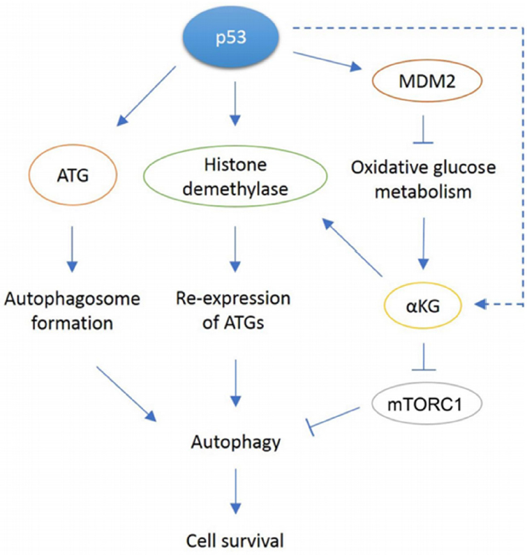
Schematic and experimental evidence for p53-driven activation of autophagy and the role of DRAM1 (open-access review and experimental studies).
03

Organelle contacts and calcium homeostasis: lysosome–ER communication

What are the molecular tethers that connect lysosomes and the ER, and how does p53/DRAM1 activity alter calcium signalling and ER stress?

Inter-organelle contact sites are key signalling hubs. Recent evidence links DRAM1 to changes in ER architecture and to modulation of STIM1-dependent calcium handling, suggesting that p53-targets can reshape intracellular signalling hubs. Mapping molecular players at these contacts and the functional consequences for calcium homeostasis will reveal new links between organelle dynamics and stress adaptation.

This programme will use split‑fluorescent reporters and proximity-labelling approaches to identify contact-site partners, combined with calcium imaging and ER stress readouts. Perturbation of candidate interactors will reveal causal steps between contact-site modulation and cellular outcomes in stress models.

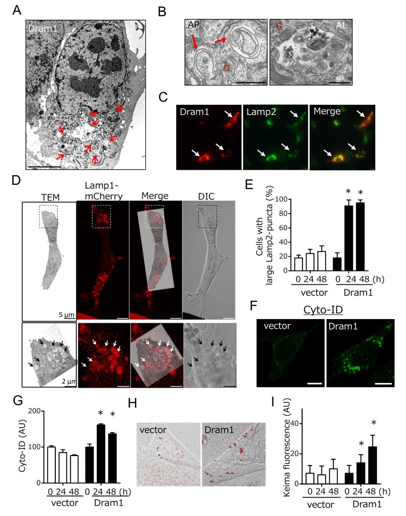
Figure(s) from Nagata et al., 2015 (PMC4502683) showing DRAM1 subcellular localization to lysosomes and effects on autophagy — relevant to lysosome–ER contact and DRAM1-driven changes in organelle structure and calcium/ER stress biology.
04

Cell-cycle control and stress-response signalling downstream of p53

How do p53-regulated transcriptional programmes intersect with cell-cycle checkpoints and proteostasis pathways to determine proliferative decisions under stress?

p53 is a master regulator of cell-cycle arrest and DNA-damage responses. Beyond canonical checkpoint enforcement, p53 targets influence proteostasis and translational quality control, which in turn feed back on cell-cycle decisions. Recent work co‑authored by Suman Rimal explores ribosome-associated quality control and stalled translation as vulnerabilities tied to stress responses.

This programme studies how p53-dependent transcriptional changes alter translation quality control, ubiquitin–proteasome and autophagy pathways to enforce cell-cycle outcomes. Combining ribosome profiling, translational stress models and cell-cycle assays will allow dissection of these integrated networks.

Figure 6 from: "The p53 target DRAM1 modulates calcium homeostasis and ER stress by promoting contact between lysosomes and the ER through STIM1" (PNAS 2024). Shows DRAM1 perturbation of Ca2+ homeostasis and DRAM1–STIM1 interaction (confocal images, co‑IP and Fluo‑2 Ca2+ imaging) relevant to p53 downstream control of organelle contact sites, calcium signaling and stress responses.